How does skin plumpness lose to return a responsibility?
summary
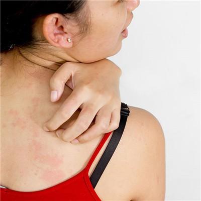
The loss of subcutaneous fat due to anorexia and malnutrition, skin aging, skin thickness thinning and elastic fiber degeneration, resulting in loss of skin fullness. Atrophic skin disease is more common in the elderly, male than female. It mainly occurs in the face, trunk and limbs. The skin showed diffuse flushing, edema, telangiectasia, skin atrophy, pigmentation and hypopigmentation. As the skin atrophy, the skin appears dry, there are chaff like scales, the skin is cigarette paper thin. This disease is seen in patients with dermatomyositis, showing skin heterochromatism, called skin heterochromatism dermatomyositis, more common in children. It may also be the early stage of mycosis fungoides, and its further development is manifested in mycosis fungoides. Progressive idiopathic skin atrophy, also known as macular atrophy: early damage for different sizes of round or irregular light red patches, and then gradually atrophy, skin color or bluish white, dark red, slightly concave or convex, wrinkled surface, feel not hard. Scleroderma and scleroderma after atrophy is different, painless itching. In the early stage, there is no feeling, generally it affects the aesthetic feeling of the appearance before seeing a doctor, and a few can cause limb disability. Most of them are young and middle-aged, especially women.
How does skin plumpness lose to return a responsibility?
The loss of subcutaneous fat due to anorexia and malnutrition, skin aging, skin thickness thinning and elastic fiber degeneration, resulting in loss of skin fullness. It is not an independent disease, but the clinical manifestations of some different diseases at a certain stage, characterized by mixed pigmentation and hypopigmentation, telangiectasia and progressive skin atrophy. Progressive idiopathic skin atrophy is a kind of skin atrophy of unknown cause, and it is progressive. It belongs to atrophic skin disease.

Dermatology: five pigmented patches of different sizes were found on the left upper limb and head and face, with a range of 1 cm × 2cm to 11cm × There was no abnormal sensation. Laboratory examination: blood and urine routine were normal, ana1: 1280. Histopathological examination: epidermis atrophied, dermal collagen fibers were loose, and skin appendages existed. The diagnosis was progressive idiopathic skin atrophy.

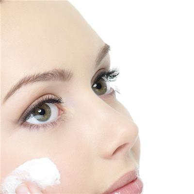
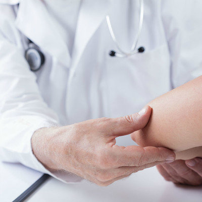
In general, western medicine has no special treatment for the disease, and most of the patients are given vitamin drugs or topical retinoic acid. According to the clinical manifestations, TCM can differentiate into deficiency of Qi and blood, liver depression and qi stagnation, yin deficiency and blood heat, spleen and kidney yang deficiency and other types, which can be treated symptomatically. Poor skin elasticity: cell elasticity atrophy, skin aging and other skin problems. Touch with your fingers, it will spring up quickly, and it will have good elasticity. Also, you can see whether the lines on your face come out. The skin with good elasticity is very smooth and tight.
matters needing attention
Patients with vascular atrophic skin heterochromatism should pay attention to the protection of the skin and avoid sunlight. Patients should be examined regularly by dermatologists to prevent malignant diseases. The skin can only be treated with protective treatment, oral Yikangning, compound vitamins, trace elements tablets, topical moisturizing cream.