How to treat adrenal cyst
summary
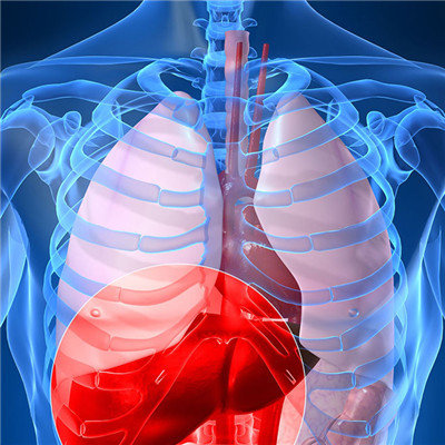
Adrenal cyst is still more common in life, we do not have to worry about this symptom. After intravenous injection of iohexol 300mg (I) ml through forearm, the scan was performed in 25 seconds, 60 seconds, 180 seconds and 360 seconds respectively. The results showed that there was an oval low-density shadow in the right adrenal area, and there was a strip-shaped dense focus with the size of about 4.8cm * 4.0cm, and no enhancement was found. The liver was enlarged and the enhancement density was normal. The wall of gallbladder was reduced, nodular and enhanced. The spleen was enlarged, and there were small pieces of low-density shadow without enhancement. The pancreas, kidney and left adrenal gland showed no abnormal enhancement. Let's share some experience.
How to treat adrenal cyst
First, adrenal cysts are mostly benign, and rarely have endocrine function. The treatment is mainly based on the patient's symptoms, cyst size and pathological changes. Cysts less than 3 cm, without clinical symptoms or endocrine function should not be treated.
Second: ① there were mass compression symptoms, cyst diameter > 5cm. ② Once hydatid cyst and neoplastic cyst are found, early surgical treatment is needed. ③ Simple cysts less than 4cm in diameter without symptoms can be observed continuously in clinic. If the cysts are enlarged or have symptoms, the operation can be performed again. ④ Functional adrenal cyst. ⑤ Malignant cyst is suspected.
Third: ① simple cyst has a complete capsule, only need to remove the cyst, keep the normal adrenal gland. There were 6 cases in this group. ② In the case of neoplastic cyst, the adrenal gland and cyst can be removed as a whole. There were 4 cases in this group. ③ For hydatid cyst, after the surrounding tissues were well protected, puncture was performed to confirm the diagnosis. After a certain amount of cyst fluid was sucked out, 4% formaldehyde solution was injected. After killing the inner head segment of the cyst, the outer cyst was cut to remove the daughter and grandson cysts. Then the inner wall of the outer cyst was wiped with formaldehyde gauze ball, and most of the outer cyst wall was cut off, and the cyst cavity was sutured layer by layer from the bottom to the outside. There was no such case in this group. ④ Most of the hemorrhagic pseudocysts were larger. When the pseudocysts could not be separated from the surrounding areas, most of the free cysts were removed, and the remaining cysts were sutured with intestinal suture.
matters needing attention
B-ultrasound showed that the adrenal gland was a smooth edge of anechoic cystic mass. CT findings: cystic mass showed thin wall, smooth edge of homogeneous water density characteristics, and can show the cyst attached to the adrenal gland, cyst wall calcification is also easy to find. MR findings: cysts were characterized by long T1 and long T2 signals with uniform intensity, and the location was more accurate.