Symptoms of white atrophy
summary
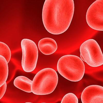
Segmental hyalinizing vasculitis is also known as atrophie blanche and livedoid vasculitis, but it is still controversial. In 1929, Milian first described the disease. Clinically, it is characterized by purpura and necrosis of the lower leg and ankle, and ivory atrophic spots after healing.
Symptoms of white atrophy
It is more common in young and middle-aged women. The lesions initially appeared as light red or bright red spots with significant pain, and further developed into purpura, which was distributed in a ring, or fused into a large dark purplish red patch on the finger cap.

Blisters may appear in the center of the lesions, and ulcers may be formed after the blisters are broken. Ulcers vary in size, small such as the size of soybeans, the big can have a dime size or even larger. The ulcer healed slowly, with typical Ivory scar, telangiectasia, purpura and pigmentation around.

Most of them occurred in the lower part of the leg, medial and lateral malleolus and around them, and a few cases involved the knee and upper limbs. The skin lesions occurred repeatedly and the course of disease was chronic without systemic symptoms.

matters needing attention
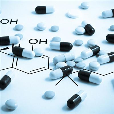
Intralesional injection of corticosteroids such as triamcinolone acetonide. Low molecular dextran, anticoagulant, ganglion blocker, nicotinic acid, SULFAPYRIDINE and so on are effective for active lesions, and can prevent recurrence. Stanozol and danazol can also be used to increase the dissolution of fibrin. Dipyridamole, enteric coated aspirin, vitamin E, Tripterygium Wilfordii and Salvia miltiorrhiza also have certain curative effect on the disease.