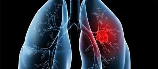
Will macular choroidal atrophy continue to develop?
summary
Will macular choroidal atrophy continue to develop? Choroidal atrophy of macular area, first of all to distinguish what causes. Choroidal atrophy, if it is pathological myopia, if the axis is still growing, then atrophy, will continue to develop. If the injury is caused by traumatic ischemia, it will not continue to develop after it has been stabilized.
Will macular choroidal atrophy continue to develop?
Choroidal atrophy is divided into primary and secondary. Primary choroidal atrophy was formerly known as chorosclerosis. It was named after the Yellow choroidal vessels under the atrophy of retinal pigment epithelium. Later, it was found that there was no pathological sclerosis in these vessels. Fundus fluorescein angiography showed that the vessels were completely filled, so it was changed to the current name.

Diffuse choroidal atrophy (DCA) 1) the age of onset is mostly 20-40 years old, and the disease gradually worsens with age, and develops into DCA around 50 years old. 2) At first, the fundus was mottled, pigment distribution was disordered, yellow dots and edema like appearance appeared, which looked like inflammation; Retinal pigment epithelium atrophy, fundus leopard shaped, choroidal vessels become thick, such as reticular. Its blood vessel wall is thickened, the central blood flow is small, and it is grayish white. Part of the blood vessels appeared to be occlusive, forming a white band.

Atrophy of the peripapillary and central choroid, loss of central vision, and susceptibility to the disease are more frequent in elderly people over 65 years of age. Incidence rate increases with age. Central localized choroidal atrophy is more common in the elderly about 60 years old, male is more than female. The early symptoms were visual distortion, central scotoma in visual field, slow progression of disease, and severe visual impairment in late stage( 3) In the late stage, the macula showed local atrophy.

matters needing attention
There was no abnormality in fundus fluorescein angiography, filling time of retinal arteriovenous fluorescein and background fluorescence outside the lesion. The intensity of fluorescence in the lesion area was alternating. Weak fluorescence showed loss of pigment epithelium and no perfusion of choroidal capillaries, while strong fluorescence showed fluorescence filling of choroidal vessels.