What does arachnoid operation preoperative examination have?
summary
Subarachnoid hemorrhage (SHA) refers to the sudden rupture of cerebral vessels caused by various reasons, but in life, many people do not know much about this early symptom, and the disease is quite serious when it comes on. Blood flow to the subarachnoid space. It is not a disease, but the clinical manifestations of some diseases, 70% - 80% of which are surgical diseases. The clinical manifestations are: what are the preoperative examinations of subarachnoid surgery?.
What does arachnoid operation preoperative examination have?
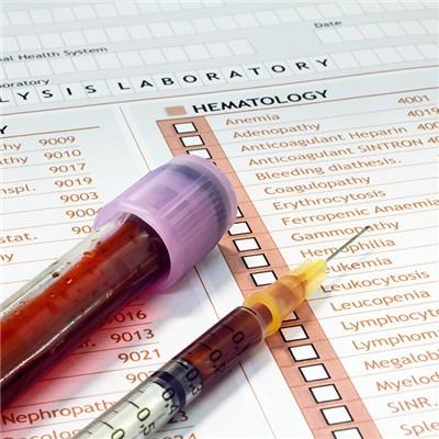
First: blood routine, urine routine and blood glucose in patients with severe cerebral subarachnoid hemorrhage in the acute phase of blood routine examination can see increased white blood cells, urine sugar and urine protein can be positive, the acute phase of increased blood glucose is caused by stress response, blood glucose not only directly reflects the metabolic state of the body, but also reflects the severity of the disease, the higher the blood glucose, stress ulcer, The higher the incidence of metabolic acidosis, azotemia and other complications, the worse the prognosis.

Second, the cerebrospinal fluid with uniform blood is the main index for diagnosing subarachnoid hemorrhage. Pay attention to lumbar puncture immediately after the onset of the disease. Because the blood has not yet entered the subarachnoid space, the cerebrospinal fluid is often negative. When the patient has obvious meningeal irritation sign, or after a few hours, the positive rate of lumbar puncture will be significantly increased, and the cerebrospinal fluid shows uniform blood, No clot.

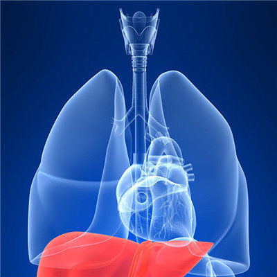
Third: imaging examination: brain CT scan or MRI examination: CT examination is the first choice for clinical suspected SAH, which is safe, sensitive and can be diagnosed early. It has high sensitivity on the day of bleeding, and can detect more than 90% SAH, showing high-density hemorrhage signs of lateral fissure cistern, anterior longitudinal fissure cistern, suprasellar cistern, cerebellopontine angle cistern, annular cistern and posterior longitudinal fissure cistern, and can determine intracerebral hemorrhage or intraventricular hemorrhage, With hydrocephalus or cerebral infarction, the condition can be observed dynamically. Most arteriovenous malformations and large aneurysms can be found by enhanced CT. Small arteriovenous malformations in the brain stem can be detected by MRI. However, it should be noted that MRI examination may induce rebleeding in the acute stage of SAH. CT can show only a small amount of hemorrhage in the midbrain cistern in about 15% of patients, which is called non aneurysmal SAH (Na SAH).
matters needing attention
One of the following cases can be examined by brain CT or MRI to prompt or exclude the disease: 1. (2) patients with history of partial epilepsy or generalized epilepsy. (3) patients with a history of chronic paroxysmal or progressive neurological dysfunction, such as hemimotor or sensory dysfunction. (4) have a history of chronic headache, other reasons are difficult to explain.