What are the symptoms of hydronephrosis in children
summary
Patients with hydronephrosis have no premonition before symptoms occur. They look like normal people, and may not show it until they are teenagers. Patients can't see its influence when they are young. This is because when children are young, their bodies don't have much waste to excrete. Even if their kidneys are damaged to a certain extent, it is enough. However, in adolescence, because their bodies grow rapidly, they need to excrete more waste. Injured kidneys may lead to hypertension, anemia, chronic kidney disease, or even renal failure. What are the symptoms of hydronephrosis in children.
What are the symptoms of hydronephrosis in children
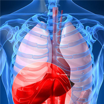
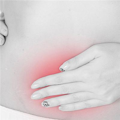
1. Clinical symptoms: most of the newborns and infants come to see a doctor with gastrointestinal discomfort and abdominal mass (accounting for more than half of them). The larger patients show intermittent lumbago and abdominal pain, hematuria, urinary tract infection and so on. Occasionally, children with renal rupture and severe hydronephrosis may have hypertension and uremia.
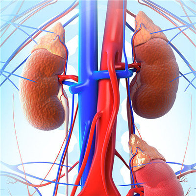
2. Make full use of biochemical, immune and other laboratory techniques in clinical diagnosis and treatment: with the improvement of various invasive detection techniques, the range of materials is expanded, and the comprehensive use of advanced and sensitive immune and molecular biology techniques shows a good application prospect in the diagnosis and treatment of hydronephrosis. Knorr et al. Used frozen sections, immunohistochemistry and RT-PCR techniques to detect the expression of nNOS in the stenotic segment of patients with congenital ureteropelvic junction obstruction (UPJO). The results showed that the nitroergic neurons and nerve fibers in the stenotic segment were significantly reduced, and the mRNA expression of nNOS was significantly decreased, It is suggested that the changes of the distribution of nitroergic nerves in UPJO stenosis may play an important role in the etiology and pathogenesis of UPJO. Jiang Lixin et al. Used S2p two-step method to detect the expression of icam21 and vcam21 in renal tissue of children with moderate and severe congenital hydronephrosis, and analyzed the relationship between them. The results showed that the expression level of icam21 in renal tissue of children with moderate and severe congenital hydronephrosis was significantly increased, It is suggested that icam21 may play an important role in the pathological development of hydronephrosis, and can be used as a clinical evaluation index of renal function damage caused by moderate and severe hydronephrosis. The results showed that ~ 22mg / Cr and ALB / Cr were the ideal indexes to evaluate the damage degree of renal tubule and glomerulus respectively, and the increase of THP / Cr and IgG / Cr in urine indicated that the renal tubule and glomerulus were seriously damaged, which was helpful to the choice of treatment methods and the prediction of curative effect in children with hydronephrosis.
3. Ultrasound has high clinical value in the diagnosis of hydronephrosis: with the rapid development of ultrasound instruments in recent years, the image is clear, the judgment is fast, and the operation of ultrasound is simple, non-invasive, no radiation and low price, so the evaluation and diagnosis of renal and urinary system abnormalities has a high position. In particular, its development does not depend on renal function, which is more valuable for patients with poor or no renal function. In children's Hospital of Chongqing Medical University, 34 cases of left hydronephrosis, 34 cases of right hydronephrosis and 25 sides of bilateral hydronephrosis were found by ultrasonography in children with urinary tract infection, hematuria or abdominal mass. The thickness of the anechoic area and renal parenchyma before and after diuresis were compared. It was found that the width and thickness of renal pelvis increased by 53% on average after furosemide administration. It was suggested that UPJ existed when the increase was 30%. ROSI et al. Suggested that diuretic B-ultrasound can make up for some deficiencies of IVU and renogram. If IVU examination indicates renal pelvis or calyceal dilatation, diuretic B-ultrasound can be used to judge whether renal pelvis dilatation is caused by obstruction or low tension of renal pelvis. Ma Hong et al. Measured the renal hemodynamic indexes of 4, JL patients with hydronephrosis, and compared them with those of healthy kidney, and concluded that hydronephrosis is a common disease. The RI of renal vessels at all levels was significantly increased, which was closely related to the degree of renal damage, especially the RI of interlobar artery (P < 0.01). Therefore, it is suggested that CDFI examination of renal blood flow, especially RI value of interlobar artery, will be helpful to make a preliminary assessment of the degree of renal function damage before operation, as well as the selection of treatment methods and the prediction of curative effect. However, it should be noted that ureteropelvic junction obstruction is often associated with other urinary malformations, up to 50% of which are reported, especially in the contralateral kidney. Therefore, when urinary malformation is found, the fetus should be checked comprehensively to prevent missed diagnosis.
matters needing attention
The main purpose of medication is to prevent and control infection before and after operation. Drugs with no damage to renal function and little damage should be used as far as possible