What examination should be done to diagnose liver cancer
summary
As we all know, liver cancer is a serious hazard, and it is likely to be life-threatening if it is not treated in time. However, many people don't know how to diagnose liver cancer, so they can't find the disease indirectly and treat it in time. Now let's take a look at what tests should be done to diagnose liver cancer.
What examination should be done to diagnose liver cancer
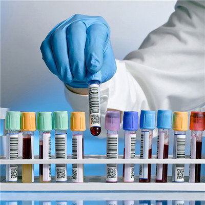
1. Blood biochemical examination: for primary liver cancer, there may be changes in liver function such as blood alkaline phosphatase, glutamic oxaloacetic transaminase, lactate dehydrogenase or bilirubin increase, albumin decrease and changes in immune indexes such as lymphocyte subsets, which is a kind of examination of liver cancer.
2. Tumor marker examination: AFP (alpha fetoprotein) is the best tumor marker in the examination of liver cancer. AFP 400 ng / ml for one month; or AFP 200 ng / ml for two months, excluding pregnancy and gonadal embryonal carcinoma, high alert to liver cancer, should be confirmed by imaging examination.
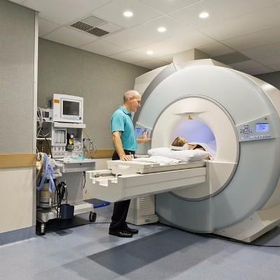
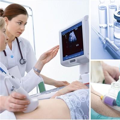
3. Imaging examination: abdominal ultrasound scanning: ultrasound scanning is noninvasive, convenient and economical, which can be used for screening high-risk groups. Intraoperative ultrasound can detect small lesions and judge the relationship between tumor and blood vessels. Ultrasound guided biopsy can directly obtain the histological diagnosis. CT examination: at present, it is the most important imaging examination method for the diagnosis and differential diagnosis of liver cancer. It is used to observe the morphology and blood supply of liver cancer, the detection, qualitative and staging of liver cancer, and the reexamination after treatment. The value of plain scan is limited, which can be used to observe steatosis, bleeding and lipiodol deposition after embolization. Enhanced scan should be regarded as routine, direct enhanced scan is feasible, and plain scan should be added according to the situation. MRI: is a powerful supplement to the imaging diagnosis of liver cancer, with the development of magnetic resonance technology more and more important. It has advantages in the detection and characterization of liver cancer under the background of fatty liver and cirrhosis, and in the judgment of tumor residue and recurrence after interventional therapy. MRI has high tissue resolution and can analyze the internal structure of the lesions. Enhanced scan can understand the blood supply of the tumor. The combination of plain scan and enhanced scan is more helpful for the diagnosis of liver cancer. Selective angiography: it used to play a key role in the evaluation of hepatocellular carcinoma, but with the clinical application of spiral CT, especially multi-slice spiral CT and MR dynamic contrast-enhanced scanning, the diagnostic value of selective angiography for hepatocellular carcinoma has gradually been replaced, and its main value at present is transarterial chemotherapy and embolization.
matters needing attention
I believe that we have a certain understanding of what examinations we should do for the diagnosis of liver cancer. When we do the above examinations, we must choose a regular hospital to ensure it. We can't choose the hospital randomly and avoid the second treatment.