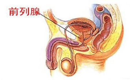
Is accessory renal artery normal?
summary
At present, retroperitoneal laparoscopic nephrectomy is the main method for the treatment of renal cell carcinoma. However, the field of vision of laparoscopic operation is small and the scope of surgical exposure is limited. Therefore, it is necessary to fully understand the relevant anatomical structure before operation, especially the blood vessels. Once damaged, it can cause massive bleeding and even endanger life. Therefore, with the increasing application of laparoscopy in the diagnosis and treatment of renal diseases, more and more attention has been paid to the research of renal vessels. So let's see if the accessory renal artery is normal??
Is accessory renal artery normal?
First, there is one renal artery on the left and one on the right. It originates from the abdominal aorta at the level of L1-2 intervertebral disc, and it flows vertically (1-2 cm below the superior mesenteric artery) into the left and right kidneys through the renal hilum. The origin of the left renal artery is often higher than that of the right renal artery. Tarzamni et al. Reported that the length of the left and right renal artery trunk was (32. 4 4 4 ± 12.0)、(35.6 ± 7) mm, the difference was not statistically significant; However, the right side of renal artery was (44. 9 9 ± 4) mm and (39. 9 7 ± 7) mm. The right renal artery was longer than the left kidney, which was confirmed by autopsy. The length of right renal artery was (46. 1 6 ± 2) mm, left (28. 6 1 ± 11.1)mm; Female right side (33.4 4 ± 1) mm, left (19. 7 1 ± 10.2)mm。
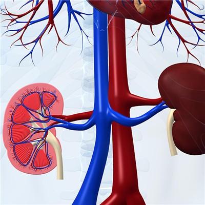
Second: under normal circumstances, with the rising position of the kidney in the embryonic period, the high blood vessels of the kidney gradually established, while the old low blood vessels gradually degenerated and disappeared. Generally, only one renal artery and vein was left. If the blood vessels were not completely degenerated, it would lead to the variation of renal blood vessels. According to the domestic literature, the total variation rate of renal artery was about 37.1%, the left side was 7.5% - 23.5%, the right side was 10.0% - 25.3%, and the bilateral side was 5.7%; The total variation rate of bilateral renal veins was about 10.2%. At present, there are many types of renal artery variation, but there is no unified view at home and abroad.
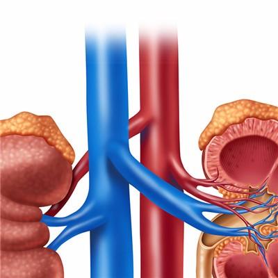
Thirdly, there are few studies on renal vein in the past. With the development of laparoscopic renal transplantation, preoperative evaluation of renal vein anatomy is becoming more and more important. It is reported that the most common variation of renal vein is multiple renal veins (2 more than 3), and most of them occur on the right side, the incidence rate is about 3%, and the incidence rate on the left side is about 1.4%. The reason may be that the right renal vein is short, close to the inferior vena cava, while the left renal vein is long, which often has complex variation.

matters needing attention
To sum up, with the continuous development of imaging technology, people have a deeper understanding of the anatomical variation of renal vessels. However, few studies from the perspective of clinical operation, that is, from the perspective of the number, concentration, angle of the renal veins into the inferior vena cava and the spatial relationship between multiple renal veins, can reflect the classification method of renal veins which is difficult to operate, Further research is needed.