Red hemangioma initial symptom?
summary
Hemangioma is a kind of congenital benign tumor or vascular malformation, which is formed by the proliferation of angioblasts during the embryonic period. It is common in the skin and soft tissue. It is more common in infants born or shortly after birth. The remaining embryonic angioblasts and active endodermoid germ invade into adjacent tissues to form endodermoid cords, which are connected with the left blood vessels after being managed to form hemangioma. The blood vessels in the tumor form their own system and are not connected with the surrounding blood vessels. Hemangiomas can occur in all parts of the body. Hemangiomas in oral and maxillofacial region account for 60% of the total hemangiomas, followed by trunk (25%) and limbs (15%). Red hemangioma initial symptom? Let's talk about it
Red hemangioma initial symptom?
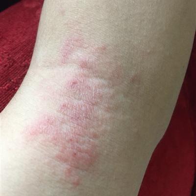
Tumor is composed of a large number of interwoven and dilated capillaries. The plaque was bright red or purplish red. It is flush or slightly raised with the skin surface, with clear boundary, irregular shape and different size. When the tumor was pressed by fingers, the color of the tumor faded; After the pressure is released, the color is restored.
Cavernous hemangioma consists of an enlarged vascular cavity and a blood sinus lined with endothelial cells. The lesions were purplish red, dark red or cyan red nodules or plaques of different sizes. The blood sinuses were of different sizes and had sponge like structures. The sinuses were filled with venous blood and communicated with each other. The surface is hemispherical or lobulated, and the volume can be reduced by pressing. Most of them are single shot. Histopathology showed that the blood vessels in the lower dermis and subcutaneous tissue expanded into irregular cavities filled with blood. The adventitia cells proliferated. It was a soft mass with no symptoms and slow growth. When the head is low, the tumor is enlarged due to hyperemia. After the body position is restored to normal, the mass will return to its original state. The superficial tumor, the surface of the skin or mucosa was purple. The skin color was normal in the deep part. When palpated, the mass was soft, with unclear boundary and no tenderness. The mass shrinks during compression and returns to its original size when the pressure is released.
The tendril hemangioma is mainly formed by anastomosing the dilated artery and vein. The tumor was like a rosary or earthworm. The palpation has the feeling of palpitation and tremor, and the auscultation has the whistling murmur. If all the feeding arteries were closed, the above pulsation and murmur disappeared.
matters needing attention
At present, drug therapy, laser therapy and surgical treatment are commonly used in the treatment of hemangioma. There is no one method to treat all types of hemangioma. It should be determined according to the type, location, depth and age of the patient. The commonly used methods are: surgical resection, radiotherapy, cryosurgery, sclerosing agent injection and laser irradiation.