Congenital cardiopulmonary hypertension?
summary
Congenital heart disease (also known as congenital heart disease) refers to the abnormal development of heart and large blood vessels at birth, which is divided into simple and complex congenital heart disease. Because the pathogenesis of complex congenital heart disease is too complex, and there are many kinds, here only introduces the relationship between several simple congenital heart disease and pulmonary hypertension. Congenital cardiopulmonary hypertension? I'd like to share my views with you.
Congenital cardiopulmonary hypertension?
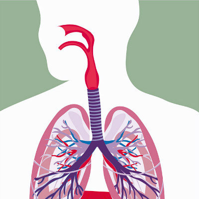
Atrial septal defect: an atrial septal defect is a hole in the septum that separates two atria. The existence of this orifice will make the arterial blood flow from the left atrium flow into the right atrium through the orifice and increase the pulmonary blood flow (see the figure below). The excessive increase of pulmonary blood flow may damage the pulmonary artery intima and cause pulmonary hypertension. Some scholars believe that atrial septal defect does not lead to pulmonary hypertension, atrial septal defect with pulmonary hypertension may be patients with idiopathic pulmonary hypertension, even without atrial septal defect, pulmonary hypertension will occur, but there is not enough evidence to support this view.

Patent ductus arteriosus: the fetus receives oxygen from the mother's lungs through the ductus arteriosus. The catheter normally closes automatically after the first cry of the baby. If the catheter does not close automatically, blood can flow from the aorta into the pulmonary artery, resulting in excessive pulmonary blood flow (see figure below), and eventually leading to small pulmonary artery injury and pulmonary hypertension. Patent ductus arteriosus is more common in preterm infants, high altitude infants and infants born to mothers infected with rubella virus in early pregnancy.
Ventricular septal defect: ventricular septal defect refers to a hole in the septum between two ventricles, which is the most common cardiac malformation. Ventricular septal defect will lead to a large amount of blood flow from the left ventricle to the right ventricle, and then into the pulmonary artery. The pulmonary blood flow will increase significantly (see the figure below), and eventually cause small pulmonary artery injury and pulmonary hypertension. If ventricular septal defect is not repaired in time, the age of pulmonary hypertension is generally younger, and there are few cases of recurrence in adulthood. The probability of pulmonary hypertension is higher than that of atrial septal defect. Studies have shown that the onset age of ventricular septal defect with pulmonary hypertension is related to the size of defect hole. The larger the defect hole is, the earlier the onset age is. Therefore, ventricular septal defect should be repaired as soon as possible if conditions permit. If pulmonary hypertension has occurred, it is extremely difficult to repair the defect.
matters needing attention
1. Cardiac ultrasound: it is the most common diagnostic method at present. The pulmonary artery pressure can be calculated by color Doppler ultrasound, but the pulmonary vascular resistance and its recovery can not be accurately evaluated, which is an important basis to determine whether the operation is feasible. 2. Right heart catheterization: To evaluate the feasibility of operation by measuring pulmonary artery pressure, calculating pulmonary resistance and partial flow of defect. 3. Pathological examination of lung tissue: take out a piece of tissue from the lung for pathological section. However, due to the difficulty of operation and the heterogeneity of the lesions, it is difficult to define the pathological state of the whole pulmonary vessels.