How to check vulvar invasive squamous cell carcinoma?
summary
Early vulvar invasive squamous cell carcinoma, also known as early vulvar squamous cell carcinoma, is generally considered to be the further development of early vulvar invasive squamous cell carcinoma by intraepithelial tumor (VIN). According to the different etiology, it can be divided into keratinized squamous cell carcinoma, HPV associated verrucous carcinoma and basal cell like carcinoma. It is usually seen in the labia major, followed by the labia minor, clitoris and perineum. It refers to the early vulvar invasive cancer with the maximum diameter of cancer focus less than 2 cm and the depth of invasion less than or equal to 1 mm. Today, let me share with you how to check for vulvar invasive squamous cell carcinoma?.
How to check vulvar invasive squamous cell carcinoma?
First, imaging examination in order to determine the clinical stage before treatment, and to make objective treatment plan, B-ultrasound, CT, MRI and lymphography of parailiac and paraaortic lymph nodes are feasible.

Second: cystoscopy and rectoscopy. Cystoscopy and rectoscopy were performed for some advanced vulvar cancer to understand the condition of bladder and rectum.

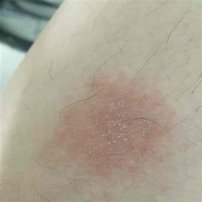
Third: all vulvar vegetations, including cauliflower foci, ulcers, nodules and white lesions, should be examined by biopsy. In order to avoid misdiagnosis, colposcopy and / or 1% toluidine blue (nuclear dye) can be used for vulvar staining, or 1% acetic acid can be used for flushing, and then biopsy can be performed. Because inflammation and cancer can show positive results, toluidine blue staining can only be used to select biopsy sites. The lesions with necrosis should be taken with enough depth, and the edge of the necrotic tissue should be taken to avoid taking only necrotic tissue and affecting the examination results.

matters needing attention
The prognosis of this disease is closely related to the size of the tumor, the depth of invasion and lymph node metastasis. The cancer with the maximum diameter ≤ 2cm, the depth of invasion ≤ 1mm and the thickness ≤ 5mm rarely has lymph node metastasis and has a good prognosis.