What are the sequelae of neonatal asphyxia?
summary
Any factor that can reduce the blood oxygen concentration can cause asphyxia. But more attention to their own body, not to check, and eventually developed into a more serious disease. Neonatal asphyxia is closely related to the intrauterine environment and delivery process of the fetus. If hypoxia occurs in the process of labor, the carbon dioxide in the fetal blood stimulates the respiratory center, resulting in strong respiratory action in the early stage. The laryngeal sphincter loses its barrier function and inhales a large amount of amniotic fluid, resulting in asphyxia during labor or neonatal asphyxia after delivery. If the fetal respiratory center has been paralyzed, then the newborn is not breathing. Today, let me talk about the sequelae of neonatal asphyxia?.
What are the sequelae of neonatal asphyxia?
First, after hypoxia, all organs can have degenerative changes, and the hypoxia susceptible areas of the brain are different at different developmental stages, so the lesion prone sites and morphology are also different. The main brain lesions are brain edema, brain tissue necrosis and intracranial hemorrhage.
Second: after necrosis, cavernous brain, polycystic brain and cortical laminar necrosis may appear. The smaller the weight of premature infants, the more vulnerable the vascular wall, the more likely to cause cerebral hemorrhage. Hemorrhage can be scattered in the ventricle, brain parenchyma, subarachnoid space and subependymal hemorrhage broken into the ventricle. Systemic blood circulation disorders lead to venous congestion, right heart enlargement, vasodilation, vascular wall permeability increase and bleeding.
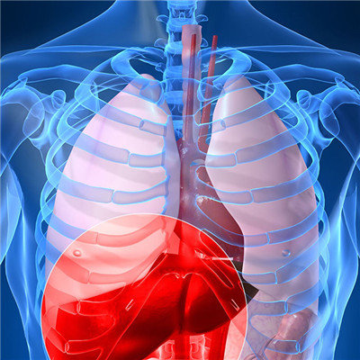
Third: full term infants with severe respiratory struggle after hypoxia, inhalation of amniotic fluid and meconium, upper respiratory tract obstruction, chest negative pressure increase, pleural heart, lung, thymus subserosal congestion bleeding correspondingly more common, respiratory system obstruction is related to the nature of inhalation. The thicker amniotic fluid and larger meconium particles are easy to stay below epiglottic cartilage, above cricoid cartilage and both sides of the bronchial orifice of the tracheal bifurcation. The thinner amniotic fluid is easy to inhale into the deep part of the respiratory tract. A large amount of keratinized epithelium or meconium particles and focal hemorrhage can be seen in the microscopic examination of lung sections. Peripheral airway obstruction may have atelectasis, incomplete obstruction may have emphysema. The patients with longer survival time may have inflammatory cell invasion, and there may be a large amount of meconium and amniotic fluid in the stomach when the gastrointestinal system is examined by naked eye. The diameter of colon is reduced and the amount of meconium is reduced.
matters needing attention
Pay attention to keep warm, try to keep the body temperature at neutral temperature about 36.5 ℃, reduce oxygen consumption. The respiration, heart sound, complexion, peripheral circulation, nerve reflex and defecation were closely observed. When breathing is stable and skin color turns red for half an hour, stop giving oxygen.