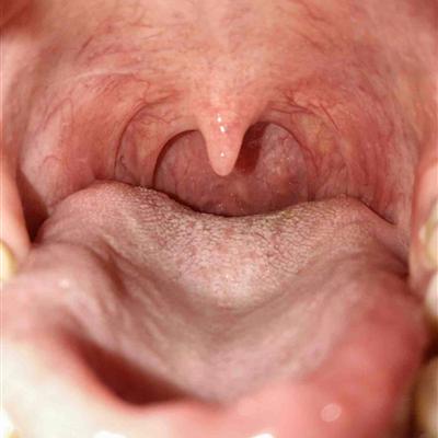
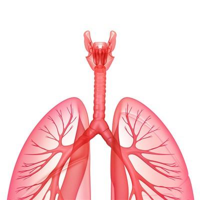
How is esophagus leukoplakia to return a responsibility?
summary
Esophageal leukoplakia is more common in men over 40 years old, generally without conscious symptoms. If the white spot is larger, thickened, accompanied by ulceration, induration, dysphagia or retrosternal pain may occur. If there is progressive dysphagia, regular reexamination should be carried out to find out if there is possibility of canceration. So let's understand how esophageal leukoplakia is going on??
How is esophagus leukoplakia to return a responsibility?
First, all the long-term persistent stimulating factors, such as strong tobacco and alcohol, spicy food and overheated diet, as well as oral hygiene, are the causes of mucosal hyperkeratosis. In addition, such as anemia, endocrine disorders, cirrhosis, systemic progressive sclerosis, fungal infection and other factors can also affect the normal keratinization process.
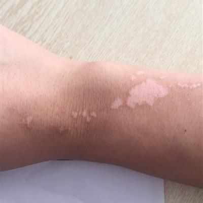
Second, hyperkeratosis and dyskeratosis of the epithelial layer, thickening of the acanthocyte layer, extensive edema inside and outside the acanthocytes, breaking of the intracellular connections, and mild infiltration of inflammatory cells in the dermis. At autopsy, there was diffuse leukoplakia in the whole esophagus, like white bark, involving the whole esophagus.

Third: or sporadic leukoplakia showed patches or plaques. Endoscopy showed that the esophageal mucosa was white or scattered white plaque, which was higher or slightly higher than the normal mucosa. Between the leukoplakia is the normal mucosa. Biopsy showed that some leukoplakia showed spinous cell thickening and contained a lot of glycogen. Therefore, this kind of mucosal leukoplakia is also called glycogenic acanthosis. Biopsy can also exclude fungi, cancer and other diseases.

matters needing attention
Esophageal leukoplakia is a rare disease. Leukoplakia can occur after keratinization of mucous membrane in various parts of the body, especially in oral and vulvar mucosa. Esophageal leukoplakia may exist alone or be a local manifestation of mucosal leukoplakia. Most of them were benign and had a good prognosis.

