How does mammary gland hyperplasia scatter calcification to return a responsibility?
summary
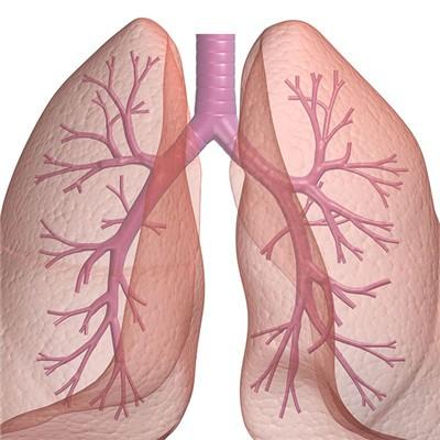
When talking about breast diseases, especially breast cancer, it is one of the diseases that threaten women's lives. Some of them even need to be removed. This will not only affect the overall aesthetic feeling, but also bring a lot of trauma to the hearts of female friends; In career, family and life to bring trouble, therefore, gynecological examination should not be underestimated, early drive out the cause, will make women's life better. How does mammary gland hyperplasia scatter calcification to return a responsibility? Let's talk about it.
How does mammary gland hyperplasia scatter calcification to return a responsibility?
There are two types of breast calcification: large calcification and micro calcification. Large calcification is usually a degenerative change in the breast, which is mostly caused by previous injury, inflammation, or aging of the breast artery, and is usually not related to cancer. Microcalcification is a calcium spot that may be found at the site where cells decompose rapidly. These residues left by rapidly decomposing cells can be shown as microcalcifications. When they appear in large groups, it means that there is a possibility of small tumors.

Clustered microcalcifications are often the only X-ray signs of early breast cancer. According to the shape, size, number and density of microcalcifications, the nature and scope of lesions can be reflected. Microcalcifications can be located in or around the mass. There are 6-15 microcalcifications with uneven density and size.

Generally speaking, breast-feeding got benign breast hyperplasia calcification, if the symptoms are not very serious, can continue to breast-feeding, but if serious, it is necessary to terminate the lactation. Treatment of mastitis, to start from cleaning the breast. Compared with benign breast hyperplasia calcification, the average density of malignant breast hyperplasia calcification group is lower, and the density and size are of great value in the differential diagnosis of benign and malignant breast diseases. The distribution of microcalcifications in mammograms seems to be irregular. However, when the cancer occurs in the terminal duct, the calcification can be located in large necrotic tissue or between cancer cells, or in the superior duct or the bifurcation of the duct or in the adjacent acinar cavity.

matters needing attention
It is necessary to remind patients to take early injection rest, suspend breast feeding, clean nipple and areola, and promote the discharge of milk. Those who need incision and drainage should stop breastfeeding. In view of the possibility of malignant transformation of lobular hyperplasia of the breast, it is suggested to improve as soon as possible without delay.