Is actinic keratosis contagious?
summary
Actinic keratosis is a kind of occupational disease, which is mainly induced by sunlight, ultraviolet rays, radioactive heat, asphalt or coal and its extractives. Lesions are more common in middle-aged men exposed to sunlight, such as face, auricle, back of hand and so on. It is mainly characterized by rough surface and keratotic scales. Remove the scales, and the base surface below is ruddy, uneven and papillary. Generally, external medicine and operation are used for treatment. Squamous cell carcinoma can be secondary in 20%. Is actinic keratosis contagious?
Is actinic keratosis contagious?
Keratinization is the process of keratin formation in the stratum corneum, which is a process of transforming cell proteins into keratins with completely different physical and chemical properties. The process of keratosis includes the fibrillation of cytoplasm and the decomposition and disappearance of cytoplasm and nucleus. Actinic keratosis is caused by long-term sunlight or ionizing radiation. It is also called actinic keratosis or senile keratosis. It is the most common epithelial precancerous skin lesion. Most of them are in the middle-aged and above men, and most of them are exposed. The main clinical manifestations were brown red or yellow flat papules or plaques. A few of them can be transformed into squamous cell carcinoma, but metastasis is extremely rare. Single cases can be local drug therapy or physical therapy. If malignant transformation is suspected, early surgical resection is recommended.

Histopathologically, it can be divided into three types: Hypertrophic type, atrophic type and carcinoma in situ type. Hypertrophic type: the hyperkeratosis of middle epidermis is obvious, and the incomplete keratosis can be seen. There are interspersed hypertrophy and atrophy of spinous layer, and the arrangement of spinous layer cells is disordered, resulting in vacuolar degeneration. Mitotic figures were common but atypical. Atrophic epidermis: atypical cells and poorly keratinized cells with loose spinous process can be seen in the basal layer. Carcinomatoid in situ: the middle epidermis is thickened, the arrangement of epidermal cells is disordered and there are atypical cells, and the boundary between epidermis and dermis is clear. There were obvious elastic degeneration in the superficial dermis of type 3, with medium density infiltration of lymphocytes.

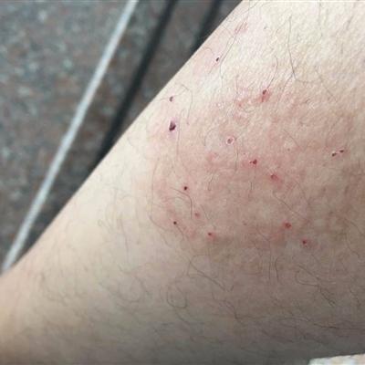
The clinical symptoms were localized brown red or yellow spots or plaques with distinct boundaries, ranging from the tip to more than 2cm in diameter, most of which were several millimeters in number. It can be slightly higher than the leather surface, but there is no obvious raised edge. The surface is rough with keratotic scales. Remove the scales by force, and you can see that the base below is ruddy, uneven and papillary. Sometimes the lesions may be angular protrusions, forming cutaneous horn lesions, which develop slowly without conscious symptoms. There may be telangiectasia around the lesion.

matters needing attention
The occurrence of this disease is closely related to sunlight exposure, especially UVB (the spectrum is 280-320nm), so those who work outdoors or often go out, it is best to apply sunscreen agents on the skin exposed to sunlight, such as 5% titanium dioxide ointment, 5% p-aminobenzoic acid ointment and sunscreen, etc. You can also take an umbrella or wear a sun hat when you go out.