Pre angiomatous symptoms?
summary
Hemangioma is a kind of congenital benign tumor or vascular malformation, which is formed by the proliferation of angioblasts during the embryonic period. It is common in the skin and soft tissue. It is more common in infants born or shortly after birth. The remaining embryonic angioblasts and active endodermoid germ invade into adjacent tissues to form endodermoid cords, which are connected with the left blood vessels after being managed to form hemangioma. The blood vessels in the tumor form their own system and are not connected with the surrounding blood vessels. Hemangiomas can occur in all parts of the body. Hemangiomas in oral and maxillofacial region account for 60% of the total hemangiomas, followed by trunk (25%) and limbs (15%). Pre angiomatous symptoms? Let's talk about it
Pre angiomatous symptoms?
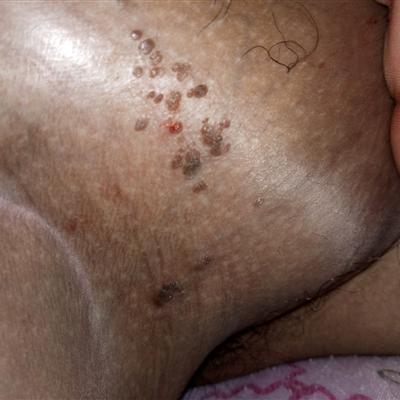
Capillary hemangioma: often occurs in the anal canal or perianal skin, also known as "wine stains." Painless, clear boundary, generally without capsule and lumen. There were a large number of well differentiated and closely arranged capillaries with thin wall, a layer of endothelial cells and basement membrane, and no smooth muscle cells. Most of them occurred in anal canal and perianal skin.
Cavernous hemangioma: common, soft as a sponge, discoloration or narrowing, vascular wall thin, flat, this type is divided into three types: ① multiple venous dilatation type. It is composed of many discrete small tumors with a diameter of less than 1 cm. ② Simple polyp type. Large volume, protruding into the intestinal cavity, often ulcers, bleeding and obstruction of the rectum symptoms. ③ Spreading and expanding type. Most of them are located in the rectum, some are single, some are multiple, the size and shape are different, and they often invade the 20 ~ 30 cm intestinal segment. Severe symptoms occurred in childhood. Under this kind of microscope, the vessels with thin wall, anastomotic, different lumen size and irregular shape can be seen, mostly in the submucosa.
Racemose hemangioma: it is composed of large caliber, thin wall, curved blood vessels, which can be veins or arteries, with arteriovenous flaccidity, and local focal mucinous degeneration. Perineum and anal canal can be seen occasionally.
matters needing attention
The principles of treatment are: 1) prevention or treatment of serious life-threatening or function related complications; ② To prevent the deformity or facial defects caused by the regression of hemangioma; ③ Prevent ulcer and infection, promote ulcer healing, reduce scar generation and relieve pain for patients with ulcer; ④ Reduce the psychological pressure of children and their families; ⑤ We should avoid over treatment for the lesions that can regress spontaneously and have a good prognosis.