Hemangioma symptom picture?
summary
When it comes to hemangioma, many people may associate it with cancer. This is not the case. Most hemangiomas are benign tumors, but it does not mean that they will not undergo malignant transformation. If hemangiomas occur in important parts of the human body, the risk is relatively high. So, what are the symptoms of hemangiomas? Let's take a look at the text.
Hemangioma symptom picture?
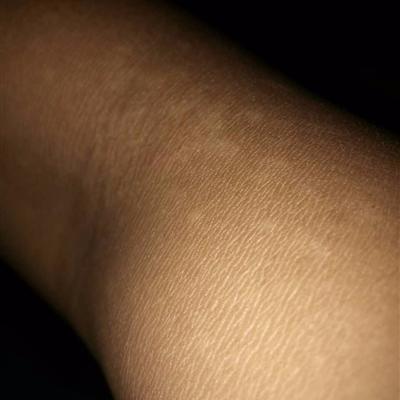
Capillary hemangioma is mostly caused by a large number of interwoven and dilated capillaries. Usually, the surface will show bright red or purple plaques. Hemangioma tends to be slightly raised, or even with normal skin. The boundary is very clear, the shape is irregular, and the size is different. When pressing with fingers, the dark color will fade, and the color will return to normal after pressure contact.
Cavernous hemangioma is composed of blood vessel lumen and blood sinuses lined with endothelial cells. The size of blood sinuses is different. Some of them are cavernous structures, so the sinuses will be filled with veins and communicate with each other. Most of the patients have no symptoms. Most of the superficial tumors are cyan purple, while the deep ones are the same as the normal skin color, And no tenderness.
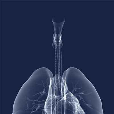
Tendril hemangioma is mainly composed of dilated arteries and veins. The high incidence of the tumor is like a rosary or an earthworm. There will be a feeling of pulsation or tremor when touching it. The wind like murmur can be heard during auscultation. If the artery supplying blood is completely occluded, the murmur and pulsation will disappear completely. Strictly speaking, the location of hemangioma is different, the symptoms are not necessary, usually we know that hemangioma occurs on the surface of the skin, but hemangioma can also occur in the human organs.
matters needing attention
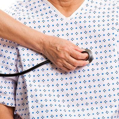
At present, drug therapy, laser therapy and surgical treatment are commonly used in the treatment of hemangioma. There is no one method to treat all types of hemangioma. It should be determined according to the type, location, depth and age of the patient. The commonly used methods are: surgical resection, radiotherapy, cryosurgery, sclerosing agent injection and laser irradiation.