Chondroma, clavicular symptoms?
summary
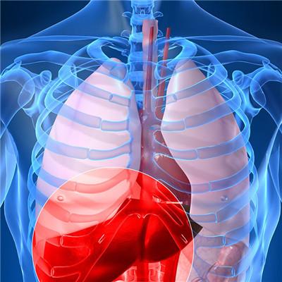
Chondrosarcoma is one of the most common malignant bone tumors, which occurs in the medullary cavity, periosteum and soft tissue. The tumors mainly occurred in the long bones of limbs and pelvis, and also in vertebrae, sacrum, clavicle, scapula and foot bone. This disease is divided into primary and secondary two kinds, the latter can be malignant from chondroma, osteochondroma, which is one of the reasons for the late onset age. This disease is common in adults, rare under 30 years old, and incidence rate increases gradually after 35 years old. Male is more than female.
Chondroma, clavicular symptoms?
In the early stage, the patient felt uncomfortable, swelling and mass appeared in a few days or weeks. In the late stage, varicose veins appeared, local skin temperature increased and congestion reddened. Patients will feel pain around the joint, initially intermittent pain, and then gradually aggravated, to persistent pain, more obvious at night, painkillers ineffective. Some patients may have joint effusion or even pathological fracture.
Primary chondrosarcoma is characterized by blunt pain, which gradually changes from intermittent to persistent, and the adjacent joints may cause limited joint movement. Local palpable mass, no obvious tenderness, surrounding skin with red heat phenomenon.
Secondary chondrosarcoma is more common in male adults over 30 years old. Most of them were in pelvis, followed by scapula, femur and humerus. Occasionally found mass, the course of disease is slow, pain is not obvious, the skin around no red heat phenomenon, near the joint can cause joint swelling, limited activity, nerve compression can cause radiation pain, numbness and so on. It is difficult to find the tumors in the chest and pelvis until they oppress the viscera and produce corresponding symptoms.
matters needing attention
1. The chondrosarcoma in medullary cavity may have patchy, wormhole like and cystic osteolytic destruction. The chondrosarcoma in medullary cavity of shaft may have large cystic bone destruction area. The inner edge of bone cortex is absorbed. When the tumor grows slowly, it can make the bone cortex thin and expand. When the bone cortex is broken, it can lead to new bone under periosteum, which is generally light. Occasionally, there is needle bone beside the cortex.